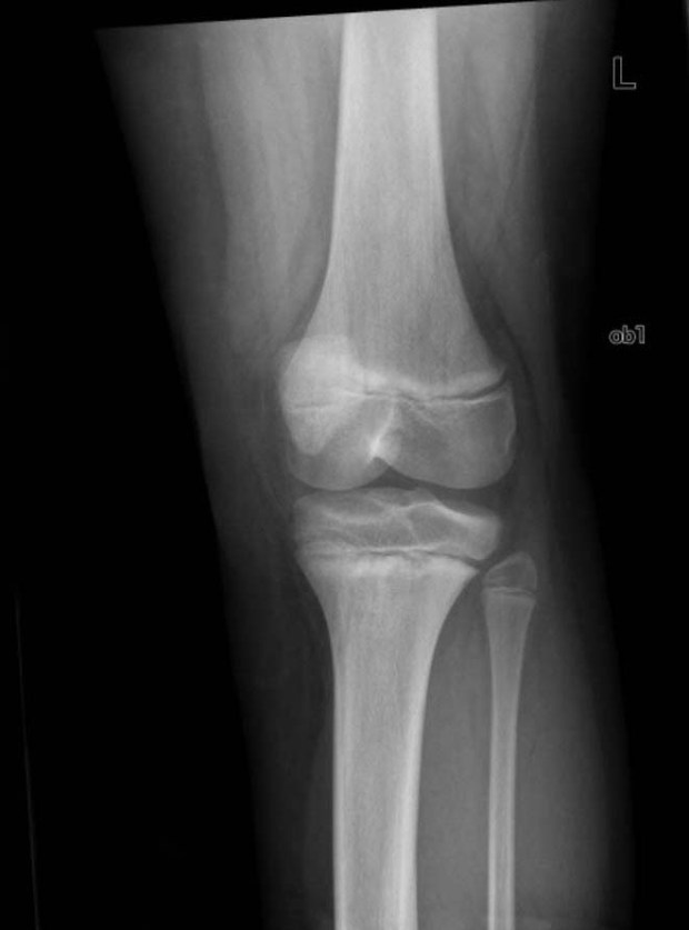
Question 1: What abnormality is seen in this picture and what exposure does it suggest?
Answer: Lead lines.
-Lead lines result from a remodeling defect of bone and are not lead deposits.
-90% of total body lead is stored in bone.
-Lead affects osteoblast and osteoclast function -> increased calcium deposition at growth plates -> Lead lines on radiographs of long bones.
-Lead lines require blood lead levels >45mcg/dL for months. Width of lines correlates with serum lead levels.
Risk factors for elevated levels in kids 1-5 years old:
- Urban dwelling
- Building before 1974
- Poverty
- Non-Hispanic black race
- High population density
– Lead is most commonly absorbed by inhalation or ingestion
– Children mainly are poisoned by paint exposure from the home
– Adults are mainly exposed in the workplace
Question 2: What are other signs of this poisoning?
Answer:
Encephalopathy
- Can develop over months with decreased memory and alertness and progress to mania or delirium
- Can be rapid in onset with seizures and coma
- Most common in toddlers: 15-30 months with blood levels >100mcg/dL
- (but has been reported with levels 70 mcg/dL or lower)
- *delayed development can occur if levels are >10 mcg/dL
GI and hematologic complaints are more common in acute poisoning
- colicky abdominal pain, metallic taste, blue-grey gingival lead lines
- constitutional symptoms
- arthralgias
*Patients may be asymptomatic even with significantly elevated levels.
*Organic poisoning: Behavioral changes, insomnia, irritability, n/v, tremor, chorea, convulsions and mania.
Question 3. What other testing should you do?
Answer: History is very important:
- Occupational exposure?
- When was the house built?
- Do they have a retained lead bullet?
- (high levels can be found years after GSW when constant exposure to bodily fluid such as CSF or synovial fluid).
- Do they have family who have had elevated lead levels?
A blood level level is the single best test.
- Levels >10mcg/dL are considered toxic by the CDC (2)
- Note: organic lead poisoning may only cause mild serum elevations, but severe elevations or urine levels.
Blood levels may take days to return so ED visit should focus on anemia and XRs for diagnosis:
Anemia
- Microcytic or normocytic
- May have evidence of hemolysis (elevated reticulocyte count)
- Basophilic stippling sometimes seen
- (this is nonspecific and seen in arsenic toxicity, sideroblastic anemia and thalassemias)
XRs
- Abdominal XR: radiopaque material in the GI tract
- Long bones (especially knee): horizontal, metaphyseal “lead lines”
Question 4: How should this patient be treated?
Answer: If lead encephalopathy is suspected -> initiate chelation therapy in the emergency department:
- Dimercaprol 75mg/m2 (or 4mg/kg IM q4 hours for 5 days)
AND, 4 hours later start:
- Edetate calcium disodium, 1500mg/m2/d IV for 5 days
Doses vary for less severe symptoms and patients who are asymptomatic
- Succimer and Penicillamine are also used.
- Succimer is oral and preferred if there is no encephalopathy because it has less side effects.
**Dimercaprol has peanut oil in the diluent, use with caution if peanut allergy.
**Do NOT confuse edetate calcium disodium with edetate disodium which is used for hypercalcemia and can lead to serious toxicity and death!
– If abdominal XR shows radiopaque flecks -> initiate whole-bowel irrigation with polyethylene glycol.
– If large lead bodies (ie. fishing sinker) -> consider surgical removal.
Disposition: Source removal!
- Do not discharge patients back to harmful environments
- Be sure to refer families and co-workers for evaluation.
Also admit:
- Children who have lead levels >70 mcg/dL or any symptoms
- Adults with neurologic symptoms
SUMMARY
- Abdominal or neurologic symptoms + hemolytic anemia -> suspicion for lead toxicity.
- Child with neurologic symptoms -> think lead toxicity!
- Emergency department diagnosis: send blood levels, but ED visit should focus on anemia and XRs for diagnosis.
- If symptomatic, initiate chelation.
Back to QUESTIONS
